Please wait...
About This Project
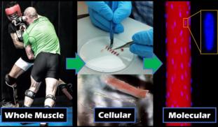
Ever wondered why some people build muscle faster than you? We know some people have more fast-twitch muscle fibers than others and muscle adaptations are regulated by your myonuclei (the cell components responsible for DNA replication, transcription, and protein synthesis). Thus, we believe the number of myonuclei differs between fiber types, and this explains much of how and why muscle grows and recovers at different rates between people, and between exercise programs!

Browse Other Projects on Experiment
Related Projects
Disrupting cancer cell signaling through drug discovery
Most cancer-related deaths are caused by metastasis, the spread of cancer cells to distant tissues. This...
CaniSense– AI-powered blood test for early cancer detection in dogs
Cancer is the leading cause of death in dogs, yet no reliable methods for early screening exist. At testblu...
Shutting down cancer’s recycling system with exosome-based therapy
Pancreatic cancer is one of the deadliest cancers because its cells survive by recycling their own components...


