Please wait...
About This Project
BBRI and Los Angeles VA Medical Center
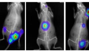
Multiple myeloma (MM) is a cancer of the bone marrow made up of plasma cells (a type of antibody-producing white blood cells). Low oxygen levels (called hypoxia) in the bone marrow increases gene expression in MM tumor cells as part of an adaptive survival response. We are targeting this hypoxic response with an experimental drug that blocks expression of these genes to ask if this molecule kills MM in the bone marrow. This is a novel approach to treat MM, an incurable disease.
More Lab Notes From This Project

Browse Other Projects on Experiment
Related Projects
Disrupting cancer cell signaling through drug discovery
Most cancer-related deaths are caused by metastasis, the spread of cancer cells to distant tissues. This...
CaniSense– AI-powered blood test for early cancer detection in dogs
Cancer is the leading cause of death in dogs, yet no reliable methods for early screening exist. At testblu...
Shutting down cancer’s recycling system with exosome-based therapy
Pancreatic cancer is one of the deadliest cancers because its cells survive by recycling their own components...


