Methods
Summary
The first step is to obtained the brain tissue, which comes to me from researchers around the world. When an animal dies of natural causes in a zoo, or in the wild, or is scheduled to be culled, researchers attempt to preserve the brain so that other researchers such as myself can study them.
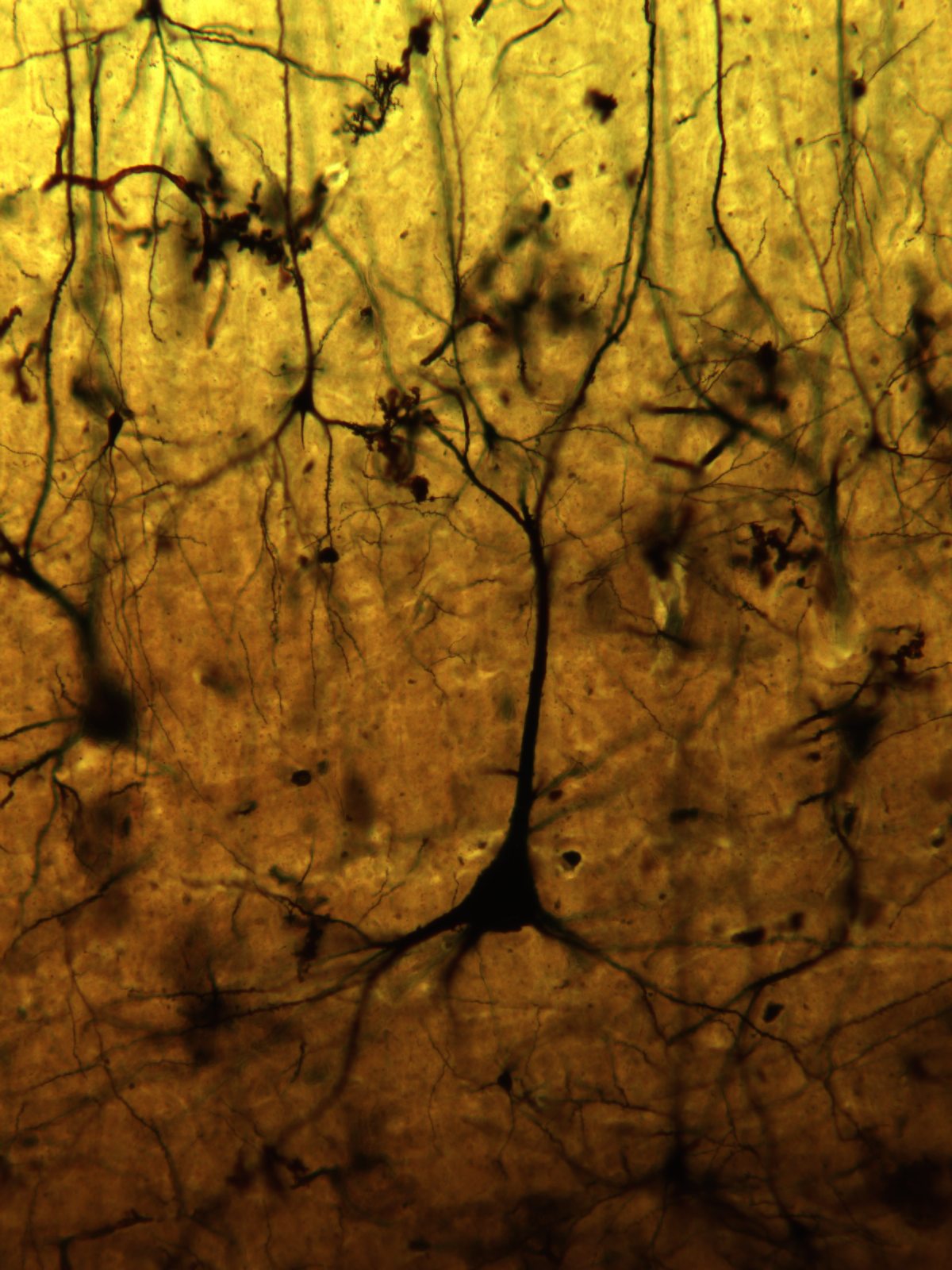
We stain the tissue with a Golgi stain, a technique that is over 100 years old, but still the gold standard for examining the morphology of neurons. Below is one such stain in a tiger:

Challenges
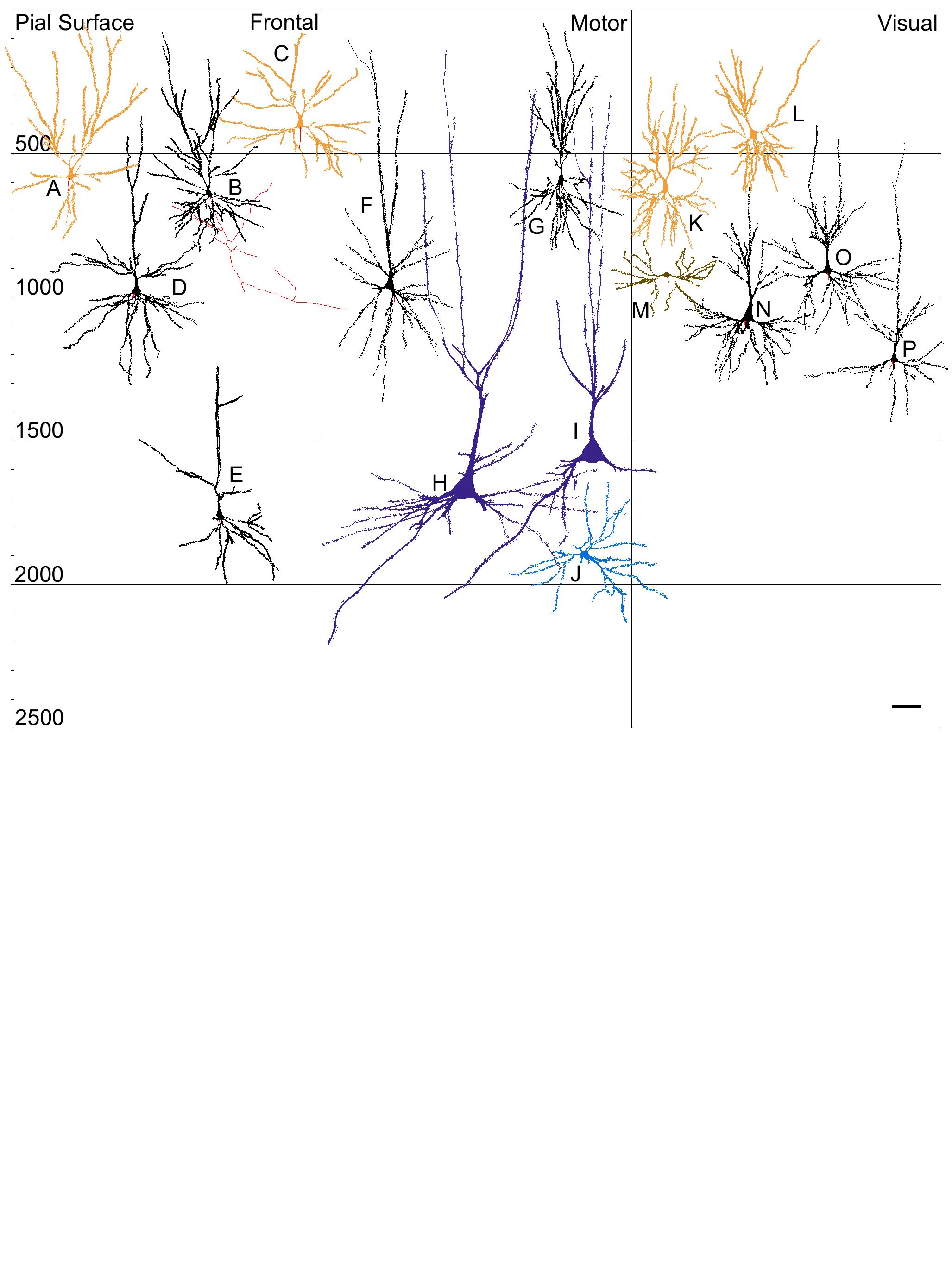
The main challenge is obtaining brain tissue from these relatively rare species. After that, the project is time consuming because it takes hours to trace each neuron. Below is an example of several neurons that have been traced in a tiger (note that neuron H is the same one in the picture above).
Note: this figure comes from (email Dr. Jacobs if you would like a pdf):
Johnson, C.B., Schall, M., Tennison, M.E., Garcia, M.E., Shea-Shumsky, N.B., Raghanti, M.A., Lewandowski, A., Bertelsen, M.F., Waller, L.C., Walsh, T., Roberts, J.F., Hof, P.R., Sherwood, C.C., Manger, P.R., & Jacobs, B. (in press). Neocortical neuronal morphology in the Siberian tiger (Panthera tigris altaica) and the clouded leopard (Neofelis nebulosa). Journal of Comparative Neurology. doi: 10.1002/cne.24022

Pre Analysis Plan
The data are examined qualitatively--note that we are seeing these neurons in these animals for the first time. The data are also examined quantitatively to see if there are significant differences among species. To do the quantitative analyses, we have a very skilled statistician who has examined many of these datasets in the past.
Protocols
This project has not yet shared any protocols.