About This Project
As corals continue to suffer from bleaching events, it is increasingly important to understand specifically how coral calcification responds on a fine-scale to bleaching. With the use of lasers in Raman Spectroscopy, this study will be the first to test how the favorability of coral skeleton formation in the wild is affected during bleaching events, which are expected to interact with the effects of ocean acidification over coming decades.
Ask the Scientists
Join The DiscussionWhat is the context of this research?
Coral skeleton growth via calcification is being affected by bleaching events. One way corals can make their growth more favorable despite unfavorable conditions is regulating the chemistry of the fluid the skeleton is made from. The extent that corals can do this over bleaching events is unknown, but dictates how well they can resist and recover from these events. This fluid is hard to study, but has been studied indirectly from coral skeleton samples called cores using lasers via a new method- Ramen Spectroscopy.
This study will use Ramen to look at how bleaching impacts corals' ability to form skeletons by looking at the high density stress bands that form in cores from bleaching. This will inform how much corals are able to be resilient amidst bleaching events.
What is the significance of this project?
This novel use of Ramen spectroscopy will be the first to analyze changes in the calcifying fluid at less than a day resolution, whereas changes have previously only been observable at a monthly resolution which do not capture bleaching events. Understanding the fine-scale responses to bleaching will help predict how reefs will change in the future with more bleaching events, as this determines their ability to grow. Having this information is critical to knowing how to best protect corals.
This project will also give insight into the processes corals use to form their skeletons, which is still debated. Not only will this increase our understanding of the organism, but also how the environment corals live in can impact their skeleton forming processes.
What are the goals of the project?
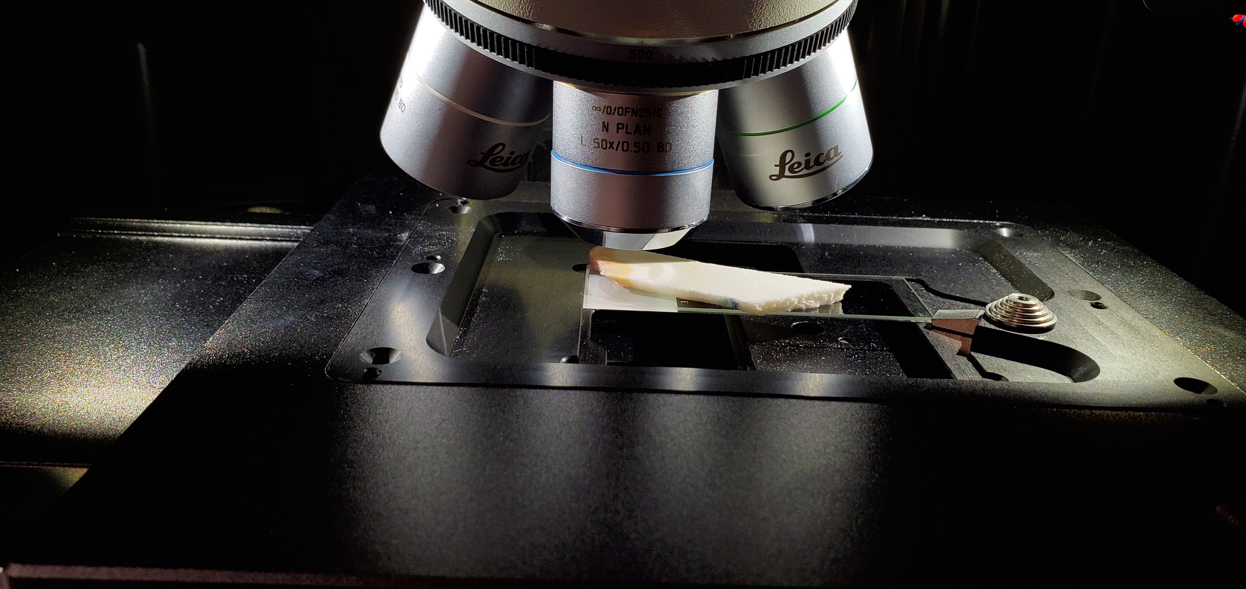
Five coral cores in Hawaii will be collected. CT scans of the cores will be taken to identify and measure bleaching stress bands within the skeletons. Cores will then be slabbed, mounted on glass slides, and sanded to a flat surface to get accurate Ramen measurements. An equation of the Ramen peak width-fluid chemistry relationship will be made using minerals formed from known conditions. Ramen spectra will be taken across the width of the stress band, representing coral growth before, during, and after bleaching. The fluid chemistry of coral cores will be calculated using the equation made from the minerals formed in known fluid conditions. Changes in the chemistry of this fluid in the cores before, during, and after bleaching will be analyzed.
Budget
To collect coral core samples, the pneumatic drill and diamond tipped core barrel will be needed. In addition, to operate the drill and support divers drilling the cores, scuba fills of compressed air will be needed. After coral cores are collected they will be slabbed using the diamond slab saw to analyze samples on the Ramen Spectroscopy. Coral slabs will be epoxied to glass slides and grinded to a smooth surface for accurate measurements. To locate stress bands in the coral cores, CT scans of the cores will be analyzed using Osiris Software.
Endorsed by
 Project Timeline
This project will be conducted Summer 2022 - April 2023. First, core cores will be collected on Oʻahu, Hawaiʻi. Core samples will then be prepared for analysis on the Raman Spectrometer. Then, methods will be tested on how to obtain the best Ramen spectra. Using those methods, a calibration curve will be created to analyze cores. Data for each replicate core will then compiled and analyzed using R and analyzed for changes in the calcifying fluid due to bleaching events.
May 23, 2022
Project Launched
Jun 12, 2022
Establish Sampling Methods
Jun 19, 2022
Create a Calibration Curve of Abiogenic Aragonite
Jul 03, 2022
Collect Coral Cores
Jul 17, 2022
Coral Core Sample Preparation
Meet the Team
Hanna Mantanona
Born on Guam and raised in Washington, Hanna completed her B.S. Marine Biology degree at Hawaiʻi Pacific University and hopes to continue her research on coral reefs as a Master's student in Marine Science. When she's not at the lab she enjoys surfing and baking sourdough bread.
Lab Notes
Nothing posted yet.
Additional Information


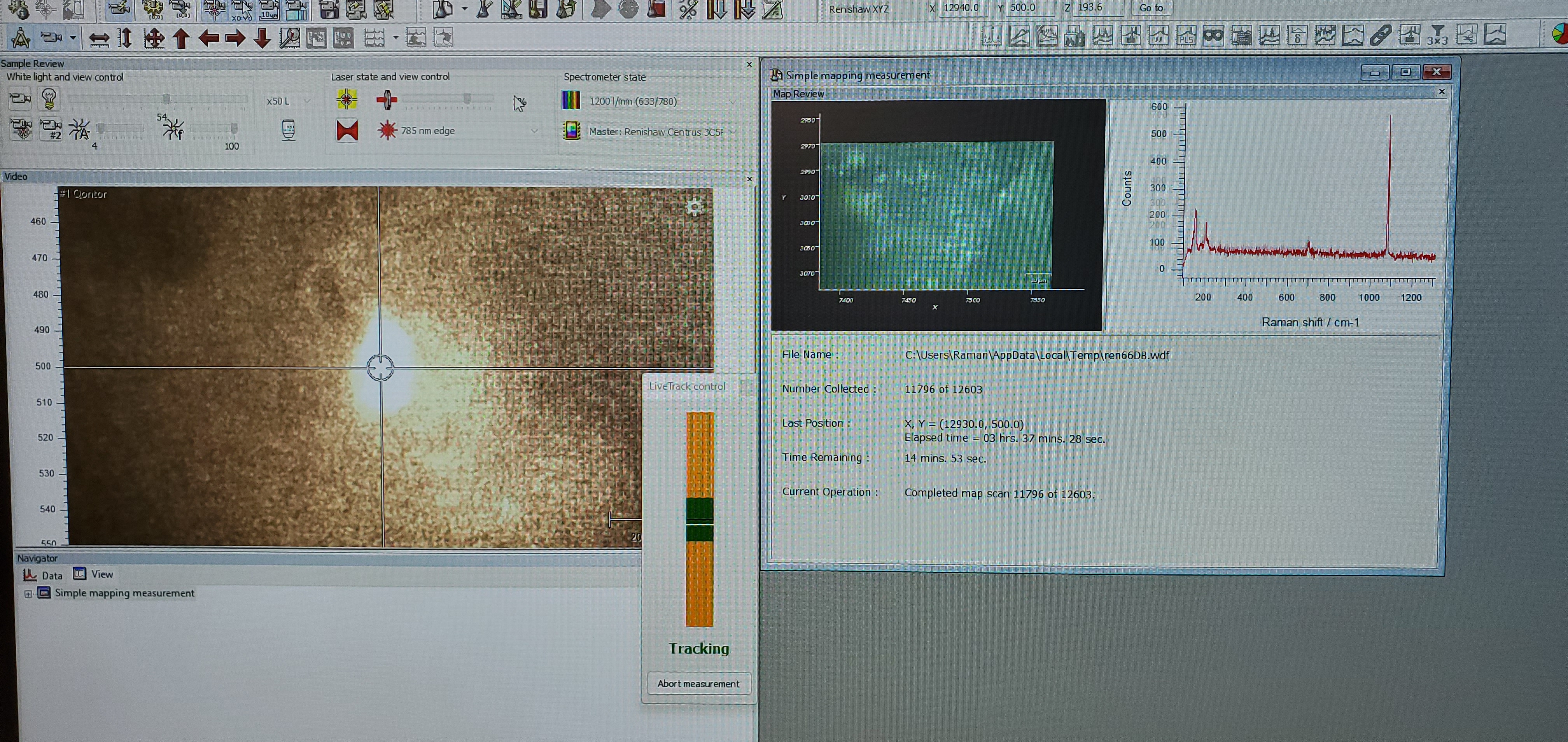
Ramen Spectroscopy works by directing a laser of a specific wavelength at a sample. When the laser hits the sample there is a shift in wavelength that is characteristic of the sample’s composition. These shifts are recorded in a graph called a spectrum. It has been found that the peak width of aragonite (the mineral coral skeletons are formed of) corresponds to the calcifying fluid saturation state. This saturation state is basically the readiness of the coral to form its skeleton. This ability for Raman Spectroscopy to study this fluid indirectly is important because the chemistry of this fluid is difficult to analyze due to it’s small volume and isolation in a few nanometers of space between the coral tissue and skeleton.
Project Backers
- 5Backers
- 63%Funded
- $3,081Total Donations
- $616.20Average Donation

