52
0
0
Like?
Please wait...
About This Project
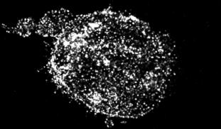
Our immune T-cells are a type of white blood cell that are crucial to recognise and attack infections and cancerous cells. For this reason, T-cells are important drug targets in vaccines and cancer immunotherapies. A drug currently in pre-clinical development is showing promising results in boosting a key regulator in T-cell functions 500-fold, but how this happens remains a mystery. We hypothesise that the drug increases the active state of the T-cell regulator by inducing a structural change.

Browse Other Projects on Experiment
Related Projects
Disrupting cancer cell signaling through drug discovery
Most cancer-related deaths are caused by metastasis, the spread of cancer cells to distant tissues. This...
CaniSense– AI-powered blood test for early cancer detection in dogs
Cancer is the leading cause of death in dogs, yet no reliable methods for early screening exist. At testblu...
Shutting down cancer’s recycling system with exosome-based therapy
Pancreatic cancer is one of the deadliest cancers because its cells survive by recycling their own components...

