Introduction to PET/CT and tumor biology
What is PET/CT, how does it work, and why is it important?
Positron emission tomography/computed tomography (better known as PET/CT) is basically a nuclear medicine technique using a device which combines a positron emission tomography (PET) scanner and an x-ray computed tomography (CT) scanner, that can combine pictures acquired from both devices into a single superposed (co-registered) image. Thus, pictures obtained by PET, which depicts the spatial distribution of metabolic or biochemical pathways measuring the uptake and distribution of specific radioactive probes and tracers in the body, can be aligned with anatomic imaging obtained by the x-ray CT scanning. Then, using powerful computer software, two- and three-dimensional reconstruction of the images can be produced and studied.
In our project, we will use these combined PET/CT images to study how different chemotherapeutic drugs alter metabolic and biochemical pathways in myeloma tumors engrafted in the bone marrow of living mice. We expect that our anti-myeloma drugs will inhibit or alter the biology of these tumor cells and this will be reflected by changes in the uptake and distribution of the various PET probes we will be studying. Many PET probes are available to study a wide range of biochemical pathways, but we will initially focus on only a few probes targeting metabolism and formation of bone lesions. For example, one probe called 18F-FDG, is commonly used to measure changes in tumor metabolism. Using PET/CT, observations that chemotherapy causes a decrease in 18F-FDG signals is indicative of tumor cell death. Therefore, using this probe, we can ask whether or not our experimental drug inhibits tumor metabolism (as a precursor to tumor death), and determine what are the kinetics (changes over time) of these effects.
PET/CT technology has revolutionized biomedical research by adding precision of anatomic localization to functional imaging, which can be performed in a non-invasive and “real time” manner. For example, we can now follow the changes in tumor and bone marrow biology and pathology from the start to the end of the study – without having to sacrifice the mice and harvest the bones mid-experiment. We can accurately measure multiple biological processes in the tumors, such as metabolism, intracellular signaling, gene expression, migration, proliferation and death using these probes and tracers. Plus, since we can visualize the tumors to specific anatomical locations in the mice (ie the bone marrow), we can study how myeloma cells behave and interact “in situ”. This makes our experimental models scientifically powerful and clinically relevant, as they very closely mirror what happens in patients with this disease.
What does the equipment look like? How do you use it? Can we see some data?
My laboratory has been able to get grants that have allowed us to purchase PET/CT and related imaging equipment. These cutting edge machines are currently being used by my laboratory and other laboratories at the Greater Los Angeles VA Healthcare system. Researchers at the VA are using PET/CT to ask important questions about cancer, brain injury, PTSD, Alzheimer's Disease, pain management, bone repair and gastro-intestinal disorders as part of the VA healthcare mission to treat our veteran population. Here are some pictures of the equipment and our facility.


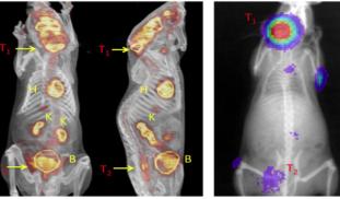
Finally, here is what our data looks like when it is first collected in our machine. Mice with tumors were imaged using the PET/CT and a radio-labeled 18F-FDG metabolic probe.

And this is what our data looks like after it has been analyzed and processed. We have been able to use our multi-modal imaging platform to identify areas were the tumors are engrafted in the bone (see the colored areas on the "luciferase images") and can cross-register those images with the uptake of the radioactive probe.

In our next set of experiments, we will treat the mice with our experimental drug and monitor the change in 18F-FDG uptake over time. The hypothesis we will be testing is whether the study drug can decrease 18F-FDG uptake compared to control animals.
1 comments